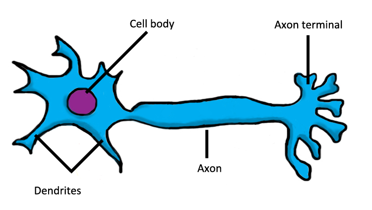

Immunolabeling of cells (immunocytochemistry or ICC) or tissues ( immunohistochemistry or IHC) with antibodies to study neurons is a highly utilized application in Neuroscience mainly due to the availability of a wide range of markers and the relatively low cost for performing and imaging the immunolabeled material. The axon hillock is located where the cell body transitions into the axon. Measurement of protein expression levelsĭownload our complimentary Neuronal Cell Markers Poster. Dendrites branch out from the cell body, where the nucleus is located.There are also axon-to-axon, dendrite-to-dendrite, and axon-to-cell body synapses. Synapses usually form between axon terminals and dendritic spines, but this is not universally true. Assessment of cellular or protein co-localization The synapse or gap is the place where information is transmitted from one neuron to another.Phenotypic and morphological analysis of neurons Example recordings of synaptic activity obtained simultaneously from the dendrite and cell body, or the cell body and axon or a layer 5 pyramidal cell are.d) axoncarry a nerve impulse away from the cell body.

a) dendritescarry a nerve impulse to the cell body b) cell bodycontains the nucleus. List the function of each part of a neuron.

microglia, astrocytes, and oligodendrocytes) a) central nervous system brain and spinal cord b) peripheral nervous system nerves.


 0 kommentar(er)
